Frequently asked questions
Heart rhythm questions, answered.
Answers to common questions about heart rhythm care, atrial fibrillation and atrial flutter. Choose a topic, then open any question to read the answer.

General heart rhythm care
What does an electrophysiologist treat?
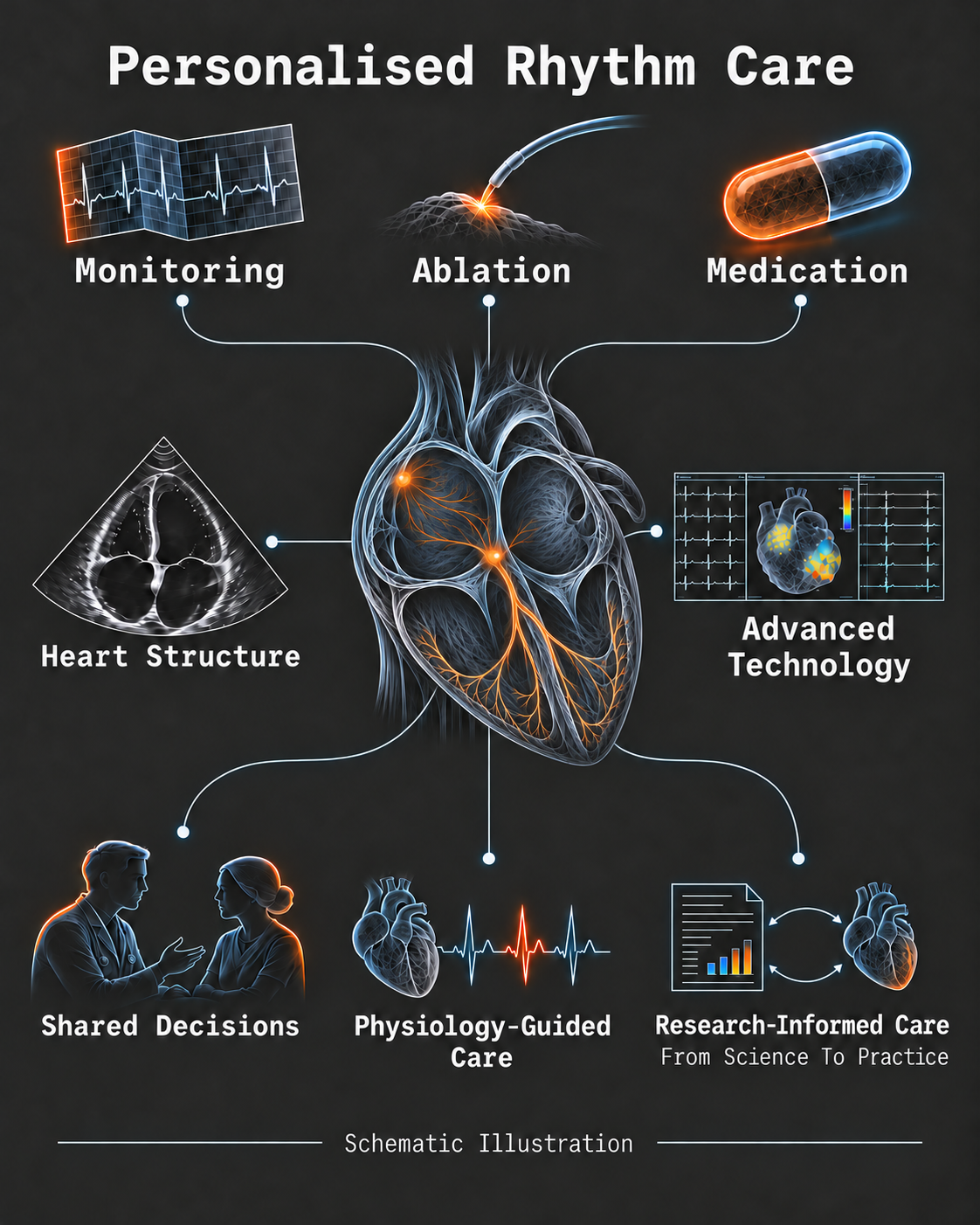
An electrophysiologist is a cardiologist who specializes in the heart's electrical system. This includes heart rhythms that are too fast, too slow or irregular, as well as some causes of unexplained fainting.
Dr. Barkagan’s practice covers the assessment and treatment of these conditions, with a particular focus on catheter ablation and implanted devices such as pacemakers and defibrillators.
When should I arrange a consultation?
A consultation can help after a new rhythm diagnosis, such as atrial fibrillation or atrial flutter, supraventricular tachycardia (SVT), premature beats (extrasystoles), or ventricular arrhythmias. It can also help with unexplained palpitations, recurrent unexplained fainting (syncope), or symptoms despite treatment.
Other reasons include side effects from rhythm medication or wanting to review a recommendation for catheter ablation or an implanted device. The purpose is to clarify the problem and discuss the options relevant to you.
What information is useful for the consultation?
Bring previous ECGs (especially ECG recordings made during arrhythmia episodes), Holter or other monitor recordings, any available heart imaging reports, and an up-to-date medication list. Relevant imaging may include an echocardiogram (heart ultrasound), cardiac CT or cardiac MRI, if these have already been performed.
Please bring copies even if the tests were done within your health fund or hospital system, as records may not be accessible across different healthcare providers in Israel. There is no need to arrange new imaging simply to prepare for the consultation unless you have been asked to do so.
These records, together with your symptoms and medical history, help determine whether further tests are needed and what they should clarify.
Does seeing an electrophysiologist mean I will need an invasive procedure?
No. Assessment comes before a treatment decision. Depending on the rhythm problem, the options may include monitoring, medication, catheter ablation or an implanted device.
The expected benefits and limitations of each option depend on your heart function, symptoms and previous treatment. Your preferences also guide the choice; a procedure is considered when it fits your individual situation.
Can I seek a second opinion about my treatment?
Yes. A second opinion can be useful after a new diagnosis or before deciding on ablation, a pacemaker or a defibrillator. It can also help when medication has not controlled the rhythm or has been difficult to tolerate.
A second opinion brings together the available recordings and previous recommendations to reassess the diagnosis, treatment goals and alternatives.
How is the choice between medication and ablation made?
The choice depends on the type of arrhythmia, its effect on your life, the condition of your heart and your preferences. Medication can help control the rate or rhythm. Ablation treats selected areas responsible for the abnormal electrical activity.
For suitable patients, ablation can be an initial option. For others, medication is a reasonable starting point or remains useful alongside a procedure. The decision is made together after reviewing your case.
For more detail, see the AF and flutter FAQ or catheter ablation FAQ.
Atrial fibrillation (AF) and atrial flutter (AFL)
What is the difference between atrial fibrillation and atrial flutter?
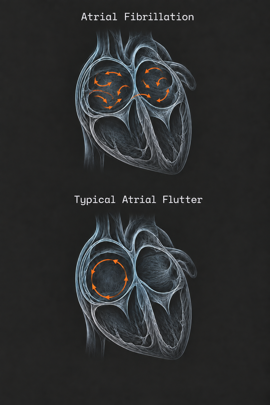
Both are rhythm disorders involving the heart's upper chambers, called the atria. In atrial fibrillation (AF), electrical activity is disorganised and the heartbeat is irregular. In atrial flutter, the electrical signal travels around an organised circuit. It’s very common for a patient to have both atrial fibrillation and flutter, or to develop another one after the initial arrhythmia has been treated. They can cause similar symptoms, but the electrical mechanism differs, and so does the treatment approach.
What causes AF?
AF is multifactorial: several factors can contribute, including heart valve disease, heart muscle disease, high blood pressure, ageing, obesity, diabetes, excess alcohol and obstructive sleep apnoea (OSA). Often there is no obvious underlying heart condition, and AF develops in a structurally normal heart. Identifying and treating contributing factors is an important part of care.1
Can I have AF without noticing it?
Yes. Some people notice palpitations, breathlessness, fatigue, reduced exercise tolerance or lightheadedness. Others have no obvious symptoms, and AF is found during an examination or rhythm recording.
How you feel is an important part of the assessment, but it does not replace checking the rhythm or assessing stroke risk.
How are atrial fibrillation and atrial flutter diagnosed?
The diagnosis requires a recording of the heart rhythm during the arrhythmia, usually an electrocardiogram (ECG). A standard 12-lead ECG taken while the arrhythmia is present can confirm it.2 When episodes come and go, a longer recording may be needed: a Holter monitor for one or several days, a patch or event monitor for several weeks, or, in selected cases, an implantable loop recorder placed under the skin for long-term monitoring.
The initial assessment usually also includes an echocardiogram (heart ultrasound) and blood tests, such as thyroid function, to look for contributing factors and to plan treatment, including stroke prevention.1
Can a smartwatch detect AF, and can I trust it?
Many smartwatches and other wearable devices can alert you to an irregular pulse, and some can also record a single-lead ECG. In a large study of more than 400,000 smartwatch users, about 0.5% received an irregular-pulse notification, and AF was found in about a third of those who later wore an ECG patch.3
A notification is a good reason to have your heart rhythm checked, but it is not a diagnosis on its own. Watches can miss AF or give false alerts, for example during movement, and they may not recognise atrial flutter when the heartbeat is regular.
The diagnosis is confirmed by a doctor who reviews an ECG recording. This can be a standard ECG or, in many cases, a single-lead ECG recorded by the device itself.2 If your watch records an ECG, save or export the tracing (for example as a PDF) and bring it to your consultation. For chest pain, fainting or severe breathlessness, seek urgent medical care rather than relying on a watch reading.
Why does it matter whether my AF comes and goes (paroxysmal AF) or is continuous (persistent AF)?
AF that stops within seven days, on its own or with treatment, is called paroxysmal AF. Persistent AF continues beyond seven days and may require treatment to restore normal rhythm. However, AF is often part of a progressive spectrum. It may begin with short episodes that become longer and more frequent over the years, sometimes progressing to a sustained form (persistent AF).
The pattern and duration of AF help guide treatment planning. Treatment results, including ablation, are better earlier in the course of the condition, while AF is still paroxysmal, than when it has been continuous for a prolonged period. Heart function and the condition of the atria also affect treatment results.
What is the difference between rate control and rhythm control?
Rate control slows the heartbeat while AF continues. Rhythm control aims to restore and maintain a normal heart rhythm (sinus rhythm), using medication, cardioversion or catheter ablation. Large clinical studies4 over the past decade have shown that early rhythm control is preferable for many patients. However, the decision should take into account the patient's heart parameters, overall health, AF duration, and individual preferences.
The approach depends on your symptoms, heart function and treatment goals. Stroke prevention is assessed separately, regardless of the approach chosen.
Do I have to try medication before AF ablation?
Not necessarily. Ablation can be considered as an initial rhythm-control treatment in suitable patients, with the aim of restoring or maintaining a normal heart rhythm over time. It is also an option when medication has not worked well or has caused side effects. In general, catheter ablation is a more targeted treatment and has been shown to be more effective than medical treatment for some patients, including those with paroxysmal AF5,6,7 and those with weakened heart muscle and heart failure8,9. Still, the decision should be personalized to each patient and their preferences.
Dr. Barkagan was also the first author of a multicentre study examining ablation as an initial treatment for persistent AF. Across nine US centres, just over half of the patients undergoing ablation had not previously tried antiarrhythmic medication. In other words, first-line ablation is already common practice in many leading centres. Rhythm outcomes after ablation were similar whether or not patients had first tried antiarrhythmic medication.10
The decision takes into account your symptoms, the type of AF, heart structure and function, previous treatment, and your values. There is no single treatment sequence that suits everyone.
How is cardioversion different from ablation?
Electrical cardioversion uses a controlled electrical shock, under brief anaesthesia, to restore normal rhythm. It does not alter the tissue responsible for the tendency to develop AF or flutter, so the rhythm can return. Cardioversion is akin to restarting the computer but it doesn’t fix it. That’s why many patients later require medication or ablation to maintain normal heart rhythm.
Ablation is a targeted treatment of the tissue that causes AF, or of the electrical pathways involved in flutter circuits. Cardioversion may be one step in a longer-term rhythm plan, whereas ablation aims to provide lasting rhythm control.
Can AF or atrial flutter be cured with ablation?
Typical atrial flutter is often treated successfully with a single ablation. Treatment interrupts the circuit in the right atrium that maintains the rhythm. Atypical flutter requires more detailed mapping because the circuit varies between patients, sometimes following previous surgery or AF ablation.
AF ablation is not a guaranteed cure, but many patients have no symptomatic AF for years after ablation. AF can recur, including without symptoms, and some patients need medication or a repeat procedure.11
Successful flutter treatment does not remove the possibility of developing AF later, and vice versa. Continued rhythm follow-up and stroke-risk assessment remain important.
Will I still need blood thinners after successful treatment?
You may. Restoring normal rhythm and preventing stroke are separate treatment decisions. Anticoagulation, often called blood-thinner treatment, is assessed according to your individual stroke risk rather than simply whether you feel well or an ablation has appeared successful.
This assessment remains relevant after treatment for either AF or atrial flutter.
References
- Joglar JA, Chung MK, Armbruster AL, Benjamin EJ, Chyou JY, Cronin EM, et al. 2023 ACC/AHA/ACCP/HRS Guideline for the Diagnosis and Management of Atrial Fibrillation: A Report of the American College of Cardiology/American Heart Association Joint Committee on Clinical Practice Guidelines. Circulation. 2024;149(1):e1-e156. doi:10.1161/cir.0000000000001193. ↩
- Van Gelder IC, Rienstra M, Bunting KV, Casado-Arroyo R, Caso V, Crijns HJGM, et al. 2024 ESC Guidelines for the management of atrial fibrillation developed in collaboration with the European Association for Cardio-Thoracic Surgery (EACTS). Eur Heart J. 2024;45(36):3314-3414. doi:10.1093/eurheartj/ehae176. ↩
- Perez MV, Mahaffey KW, Hedlin H, Rumsfeld JS, Garcia A, Ferris T, et al. Large-Scale Assessment of a Smartwatch to Identify Atrial Fibrillation. N Engl J Med. 2019;381(20):1909-1917. doi:10.1056/NEJMoa1901183. ↩
- Kirchhof P, Camm AJ, Goette A, Brandes A, Eckardt L, Elvan A, et al. Early Rhythm-Control Therapy in Patients with Atrial Fibrillation. N Engl J Med. 2020;383(14):1305-1316. doi:10.1056/nejmoa2019422. ↩
- Andrade JG, Wells GA, Deyell MW, Bennett M, Essebag V, Champagne J, et al. Cryoablation or Drug Therapy for Initial Treatment of Atrial Fibrillation. N Engl J Med. 2021;384(4):305-315. doi:10.1056/nejmoa2029980. ↩
- Wazni OM, Dandamudi G, Sood N, Hoyt R, Tyler J, Durrani S, et al. Cryoballoon Ablation as Initial Therapy for Atrial Fibrillation. N Engl J Med. 2021;384(4):316-324. doi:10.1056/nejmoa2029554. ↩
- Turagam MK, Musikantow D, Whang W, Koruth JS, Miller MA, Langan MN, et al. Assessment of Catheter Ablation or Antiarrhythmic Drugs for First-line Therapy of Atrial Fibrillation: A Meta-analysis of Randomized Clinical Trials. JAMA Cardiol. 2021;6(6):697-705. doi:10.1001/jamacardio.2021.0852. ↩
- Marrouche NF, Brachmann J, Andresen D, Siebels J, Boersma L, Jordaens L, et al. Catheter Ablation for Atrial Fibrillation with Heart Failure. N Engl J Med. 2018;378(5):417-427. doi:10.1056/nejmoa1707855. ↩
- Prabhu S, Taylor AJ, Costello BT, Kaye DM, McLellan AJA, Voskoboinik A, et al. Catheter Ablation Versus Medical Rate Control in Atrial Fibrillation and Systolic Dysfunction: The CAMERA-MRI Study. J Am Coll Cardiol. 2017;70(16):1949-1961. doi:10.1016/j.jacc.2017.08.041. ↩
- Barkagan M, Milman A, Zahavi G, Younis A, Dhakal B, Dixit S, et al. Catheter Ablation as First-Line Therapy in Persistent Atrial Fibrillation: Patient Characteristics and Clinical Outcomes. JACC Clin Electrophysiol. 2024;10(6):1078-1086. doi:10.1016/j.jacep.2024.02.035. ↩
- Andrade JG, Deyell MW, Macle L, Wells GA, Bennett M, Essebag V, et al. Progression of Atrial Fibrillation after Cryoablation or Drug Therapy. N Engl J Med. 2023;388(2):105-116. doi:10.1056/nejmoa2212540. ↩
Catheter ablation
What is catheter ablation, and which rhythm problems can it treat?
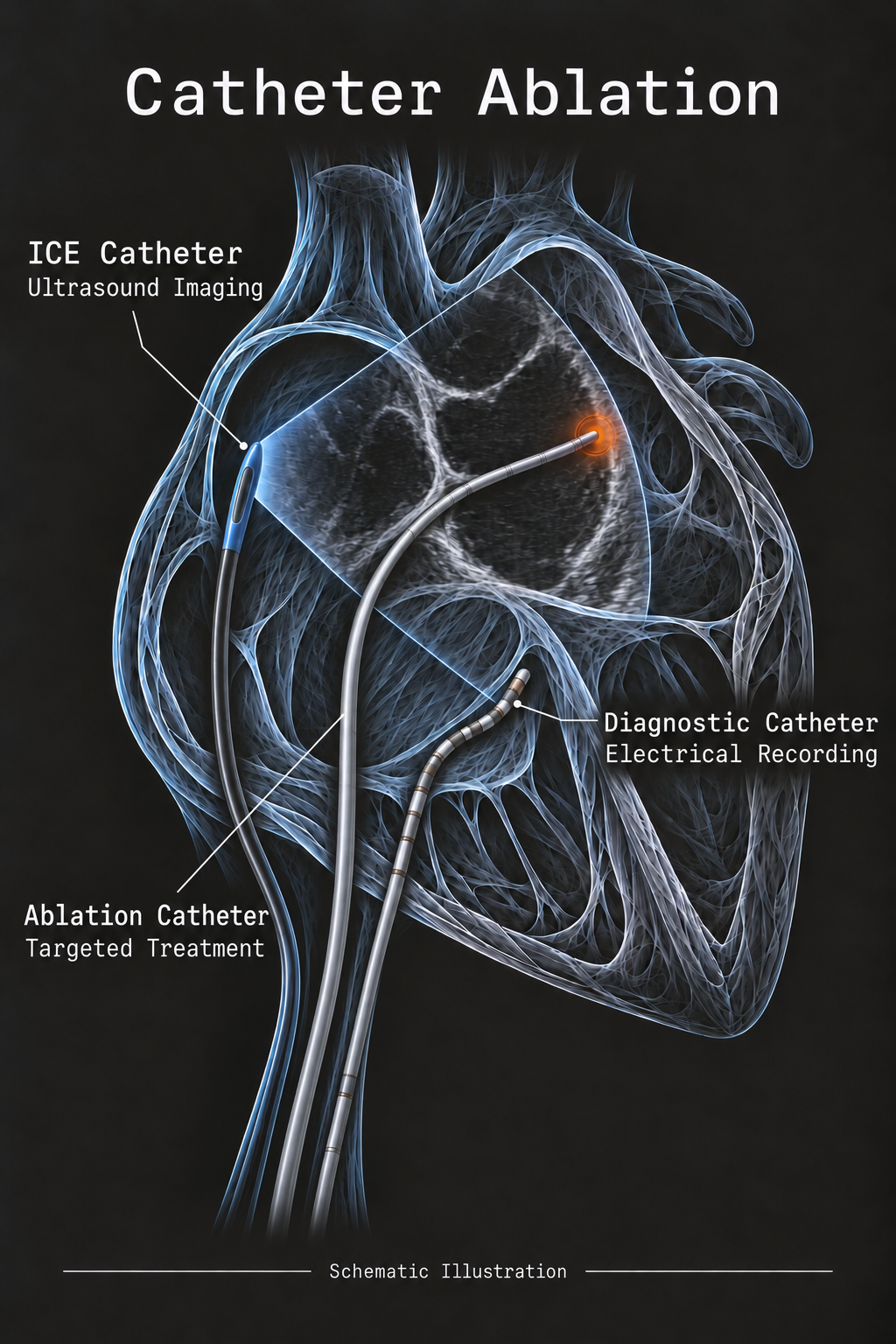
Catheter ablation treats areas of heart tissue responsible for an abnormal rhythm. It is a minimally invasive procedure in which thin catheters are guided through blood vessels, usually from the groin, and positioned in the heart. Precise recording of the heart’s electrical activity shows the mechanism of the arrhythmia, its path and the optimal treatment target. Once the target is identified, energy is applied in a focused way to interrupt abnormal signals or isolate their source.
Ablation can treat atrial fibrillation, atrial flutter, supraventricular tachycardia, and selected rhythms arising in the lower chambers, including ventricular tachycardia and troublesome premature beats. The approach differs for each condition.
How is it decided whether ablation is right for me?
The decision depends on your rhythm recordings, symptoms, heart structure and function, previous treatment and general health. The expected benefit of ablation is weighed against its risks and the alternatives.
The aim varies. For some rhythms, ablation may eliminate the problem. For others, the realistic goal is fewer episodes, less time in an abnormal rhythm or fewer defibrillator shocks.
What does ablation for atrial fibrillation involve?
The foundation is electrical isolation of the pulmonary veins. The pulmonary veins carry blood from the lungs into the upper-left chamber of the heart and are a common source of electrical signals that trigger atrial fibrillation (AF).
Ablation creates an electrical barrier around their connections to the atrium. Additional treatment may be appropriate depending on the type of AF, the atrial tissue and the electrical findings during the procedure.
What is the difference between radiofrequency, freezing and pulsed field ablation?
Radiofrequency ablation uses heat. Cryoablation uses freezing. Pulsed field ablation uses short electrical pulses to affect the targeted heart muscle cells.
Radiofrequency ablation passes an electrical current through the catheter tip and heats a small area of tissue, usually one point at a time, to create a lasting scar that blocks abnormal signals. Cryoablation cools tissue to well below freezing; for AF it is most often delivered with a balloon that surrounds the opening of each pulmonary vein. Pulsed field ablation delivers very short, high-voltage pulses that open tiny pores in the membranes of heart muscle cells, so the targeted cells stop working without significant heating. Heart muscle is more sensitive to these pulses than many neighboring tissues, such as the esophagus and nearby nerves, which is one reason for the growing use of this method. [1, 5]
In a randomized trial in paroxysmal AF, pulsed field ablation was no less effective and no less safe than thermal ablation (radiofrequency or cryoablation) at one year. [5] In a study co-authored by Dr. Barkagan, patients treated with pulsed field ablation had less chest pain from inflammation around the heart after the procedure than those treated with radiofrequency ablation. [6]
Each method has different properties and uses. The best choice depends on the arrhythmia, the tissue being treated, the procedure needed, and which options are suitable and available for your case.
How is the ablation target identified?
The target depends on the rhythm problem. Catheters record electrical signals from inside the heart, making it possible to work out where an abnormal rhythm starts or how it travels. Pacing from selected sites can help confirm the mechanism and identify the tissue that needs treatment.
For AF, the main target is usually the electrical connection between the pulmonary veins and the left atrium. For other rhythms, treatment may target a small focus, an extra electrical pathway, or a critical part of an electrical circuit. Electrical testing after ablation helps check whether the intended pathway has been blocked or the source isolated.
What kinds of catheters are used during ablation?
Several types of catheters are used in one procedure, each with a different role. Diagnostic catheters are placed at fixed positions in the heart to record and stimulate its electrical activity. Mapping catheters carry many small electrodes and, together with the mapping system, collect a large number of recordings to build a detailed electrical map of the heart chamber. Dr. Barkagan’s preclinical research in pigs has evaluated multielectrode mapping catheters with small, closely spaced electrodes, which increased the speed and detail of mapping in both the atria and the ventricles. [2, 7, 8]
An imaging catheter for intracardiac echocardiography (ICE) provides live ultrasound images from inside the heart (see question 8). The ablation catheter delivers the energy that treats the target tissue: radiofrequency, freezing or pulsed field energy, either at a single point or through larger designs such as balloons or expandable electrodes. Dr. Barkagan’s preclinical studies have also characterized new ablation catheter designs, including an expandable lattice electrode for radiofrequency and pulsed field ablation. [9, 10]
What is electroanatomic mapping, and how is the catheter navigated inside the heart?
Electroanatomic mapping combines a three-dimensional model of the heart chamber with electrical recordings from many locations. The system tracks the catheters and displays their positions on the map, helping the physician navigate to the treatment area and return to a previously identified point.
Color-coded maps show when different areas activate and the strength of their electrical signals. These findings help identify abnormal circuits and tissue affected by scarring. High-density mapping collects many closely spaced recordings to reveal details that may be difficult to distinguish on a simpler map. Mapping supports precise targeting and reduces reliance on X-rays, lowering radiation exposure. [1]
Improving the detail and accuracy of these maps is also a focus of Dr. Barkagan’s research. His preclinical work has examined how the spacing of recording electrodes affects the distinction between local electrical activity and signals from surrounding tissue, a practical question when mapping scar-related arrhythmias. [2]
What is intracardiac echocardiography (ICE), and why is it useful?
ICE is ultrasound performed using a small catheter placed inside the heart through a vein. It provides live images of the chambers, valves and catheters. While the electrical map helps explain the rhythm, ICE shows the anatomy and the relationship between the catheter and the heart tissue.
Dr. Barkagan routinely uses ICE in all left-sided ablation procedures (such as ablation for atrial fibrillation or left-atrial flutter). It helps guide access to the left atrium, assess catheter position and contact, and detect fluid accumulating around the heart early. Together with mapping, it can reduce the need for X-ray fluoroscopy, minimizing exposure to ionizing radiation. [1]
Large observational studies of AF ablation have associated ICE use with fewer complications. It provides an additional layer of guidance and safety. [3]
What preparation is needed, and will I be asleep?
Preparation includes a review of your rhythm recordings, medical history, imaging and medications. An echocardiogram is reviewed or arranged when needed; additional scans are used selectively. Medication and anesthetic instructions are confirmed for your procedure. Some patients are also referred to an anesthesiologist for evaluation before the procedure.
In Dr. Barkagan’s practice, AF ablation and most left-atrial flutter procedures are usually performed under general anesthesia. Typical right-atrial flutter and some supraventricular tachycardia procedures can be performed under sedation. The plan depends on both the procedure and your health.
Should I stop my blood thinner before ablation?
No, unless specifically instructed. For AF ablation, anticoagulation (blood-thinner treatment) is usually continued without interruption. The instructions depend on the medication and the procedure. [1]
Before AF ablation, effective anticoagulation for at least three weeks is generally recommended, and in some cases imaging is used to make sure there is no blood clot in the heart. After ablation, anticoagulation usually continues for at least two months; the long-term decision depends on your individual stroke risk, not on whether the ablation was successful. [1]
Do not stop or change a blood thinner on your own. You will receive specific instructions for your medication and your procedure.
How successful is catheter ablation?
Success depends on the rhythm being treated and the condition of your heart. Atrial flutter and many common forms of supraventricular tachycardia can often be treated successfully with one procedure.
For AF, the aim is sustained normal rhythm or a substantial reduction in episodes. For ventricular tachycardia associated with heart muscle disease, the goal is often fewer episodes and defibrillator shocks.
What are the risks of ablation?
Serious complications are uncommon in routine ablation procedures, but the risk depends on the rhythm being treated, the complexity of the procedure and your health. Studies of AF ablation have reported lower complication rates in more recent years, which may partly reflect accumulated experience and technological advances. [4]
Electroanatomic mapping improves targeting and reduces radiation exposure. ICE provides live ultrasound guidance and helps detect complications early; Dr. Barkagan routinely uses it in all left-sided procedures. These measures support safety but do not remove all risk. The decision to proceed weighs the expected benefit against the risks relevant to your case and the alternatives.
Is catheter ablation more dangerous than antiarrhythmic medication?
Not necessarily. Ablation is precise and targeted: it treats only the small areas of heart tissue responsible for the arrhythmia. Antiarrhythmic drugs change the electrical properties of heart muscle cells throughout the heart, and some also affect other organs. [11]
The risks of ablation are concentrated around the day of the procedure, and serious complications are uncommon. [4] The side effects of antiarrhythmic drugs, by contrast, can appear at any time while the medication is taken, and some, such as those of amiodarone, become more likely with long-term use. [11] In randomized trials of first-line treatment for paroxysmal AF, no statistically significant difference in serious adverse events was observed between ablation and antiarrhythmic drugs, whereas ablation was associated with fewer arrhythmia recurrences and hospital admissions. [12]
The choice between them depends on your arrhythmia, your heart condition and your preferences (see question 2).
What happens during recovery and follow-up?
After the procedure, you are monitored during recovery. An overnight hospital stay for observation is common, although the plan varies depending on the procedure and your recovery. Written discharge instructions explain activity, medication and symptoms requiring urgent contact.
Most patients return to light daily activities within a few days. Strenuous exercise and heavy lifting are usually avoided for about a week to let the puncture site in the groin heal, and many patients feel tired or have mild chest discomfort for several days. After AF ablation, the heart tissue continues to heal for about three months; brief episodes of arrhythmia during this period are common and do not necessarily mean the procedure has failed. [1, 6] The exact timing for returning to work, driving and exercise depends on the procedure and your recovery, and is explained before discharge.
Follow-up reviews symptoms, rhythm recordings and medications. A Holter or other monitor may be used to assess the rhythm over time. Rhythm medication may continue during the early healing period and be reassessed later.
Is a repeat ablation an option if the arrhythmia returns?
Yes, a repeat ablation is often a reasonable option. The first step is to identify why the arrhythmia has returned. After AF ablation, this can involve electrical reconnection of a pulmonary vein or another circuit within the atrium.
Detailed electroanatomic mapping (see question 7) guides treatment of the mechanism found. A repeat ablation is planned around those findings rather than simply repeating the previous procedure.
Selected references
- [1] 2024 EHRA/HRS/APHRS/LAHRS expert consensus statement on catheter and surgical ablation of atrial fibrillation. Read source
- [2] Barkagan M, et al. Optimizing Interelectrode Distance for Accurate Mapping of Postinfarct Scars: Insights on Electrogram Characteristics. JACC Clin Electrophysiol. 2025;11(6):1132-1141. Read source
- [3] Differences in outcomes among patients with atrial fibrillation undergoing catheter ablation with versus without intracardiac echocardiography. 2022. Read source
- [4] Procedure-Related Complications of Catheter Ablation for Atrial Fibrillation. J Am Coll Cardiol. 2023. Read source
- [5] Reddy VY, et al. Pulsed Field or Conventional Thermal Ablation for Paroxysmal Atrial Fibrillation. N Engl J Med. 2023;389(18):1660-1671. Read source
- [6] Yadin D, Barkagan M, et al. Pulsed Field Ablation Is Associated With Fewer Post-Procedural Pericardial Inflammatory Symptoms Compared With Radiofrequency Ablation. J Cardiovasc Electrophysiol. 2026;37(9):1930-1938. Read source
- [7] Barkagan M, et al. A novel multielectrode catheter for high-density ventricular mapping: electrogram characterization and utility for scar mapping. Europace. 2020;22(3):440-449. Read source
- [8] Sroubek J, Rottmann M, Barkagan M, et al. A novel octaray multielectrode catheter for high-resolution atrial mapping: Electrogram characterization and utility for mapping ablation gaps. J Cardiovasc Electrophysiol. 2019;30(5):749-757. Read source
- [9] Barkagan M, et al. Expandable Lattice Electrode Ablation Catheter: A Novel Radiofrequency Platform Allowing High Current at Low Density for Rapid, Titratable, and Durable Lesions. Circ Arrhythm Electrophysiol. 2019;12(4):e007090. Read source
- [10] Yavin H, Shapira-Daniels A, Barkagan M, et al. Pulsed Field Ablation Using a Lattice Electrode for Focal Energy Delivery: Biophysical Characterization, Lesion Durability, and Safety Evaluation. Circ Arrhythm Electrophysiol. 2020;13(6):e008580. Read source
- [11] Joglar JA, et al. 2023 ACC/AHA/ACCP/HRS Guideline for the Diagnosis and Management of Atrial Fibrillation. Circulation. 2024;149(1):e1-e156. Read source
- [12] Turagam MK, et al. Assessment of Catheter Ablation or Antiarrhythmic Drugs for First-line Therapy of Atrial Fibrillation: A Meta-analysis of Randomized Clinical Trials. JAMA Cardiol. 2021;6(6):697-705. Read source
Consultation
If you would like to review your diagnosis or treatment options, a consultation can help clarify the next step and which options fit your rhythm, heart health and priorities.